CBCT is performed using a single rotation of 360° in order to acquire the image in volume at a resolution of the highest quality and in a very short time.
Thanks to the 51 different modes of examination, NewTom VGi evo offers a highly effective tool adapted to the needs of all clinical situations. The different scan modes adapts perfectly to every anatomical region to be analyzed.Scanning surfaces (FOV) suitable for the investigation of different anatomical regions are governed by international standards according to the ALARA (As Low As Reasonably Achievable) principle, which aims at minimizing the effective dose absorbed by the patient.
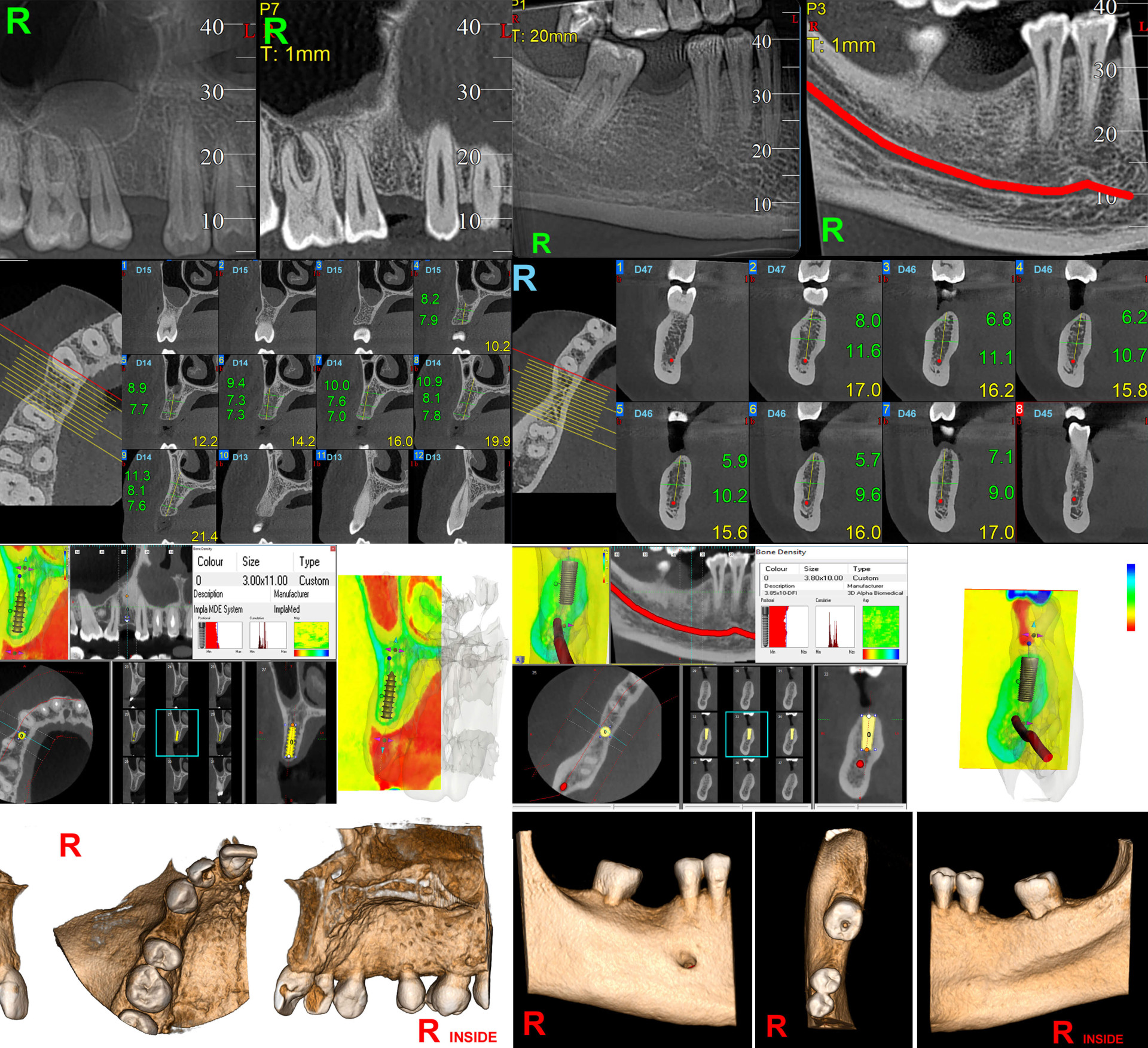
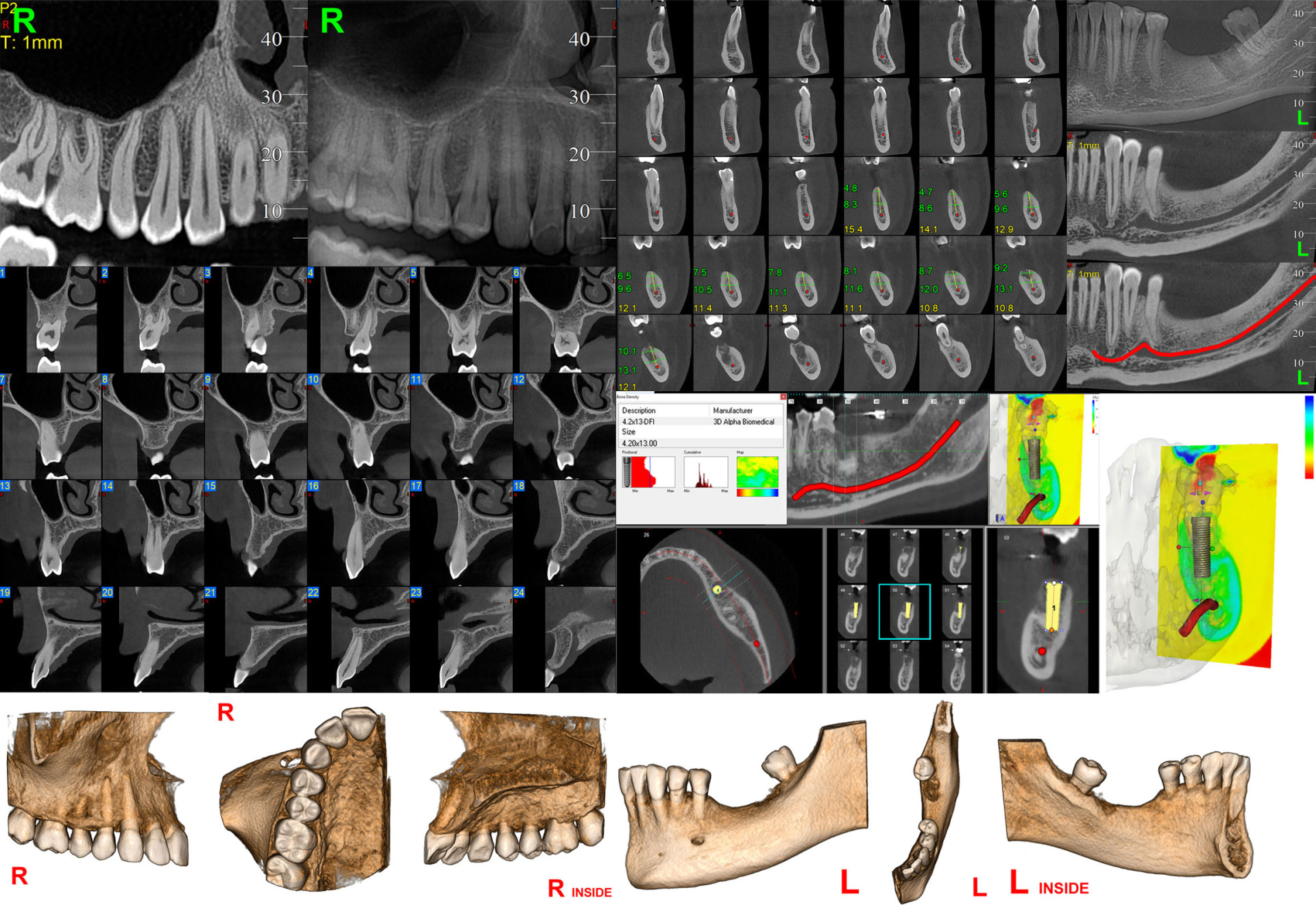
* Information on graphic elements of the tomography, bone density and implant simulations can be viewed in this INDEX.
Unidental tomography is a three-dimensional imaging technique used in dentistry, which provides accurate and detailed images of a single dental area, helping to diagnose and treatment planning in dentistry.
Hemiarcade tomography is a three-dimensional imaging technique used in dentistry, which provides accurate and detailed images of half a dental arch, providing essential information for diagnosis and treatment planning in dentistry.
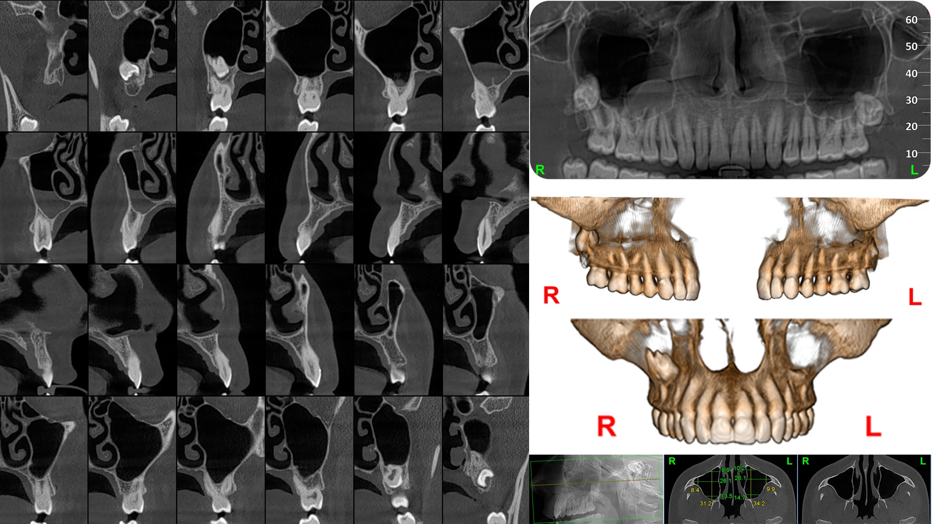
Maxillary arch tomography is a three-dimensional imaging technique used in dentistry, which provides accurate and detailed images of the maxillary arch, zygomatic arches and maxillary sinus, providing essential information for diagnosis and treatment planning in dentistry.
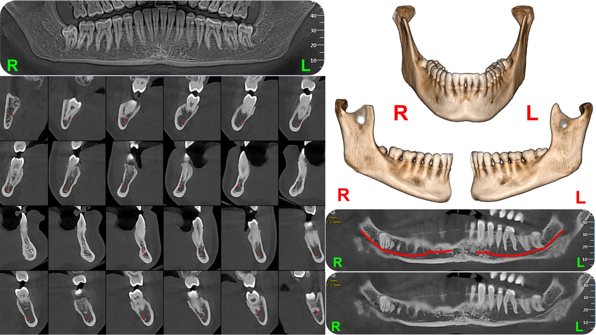
Tomography of the mandibular arch is a three-dimensional imaging technique used in dentistry, which provides accurate and detailed images of the mandibular dentoalveolar process and the mandibular canal, providing information essential for diagnosis and treatment planning in dentistry.
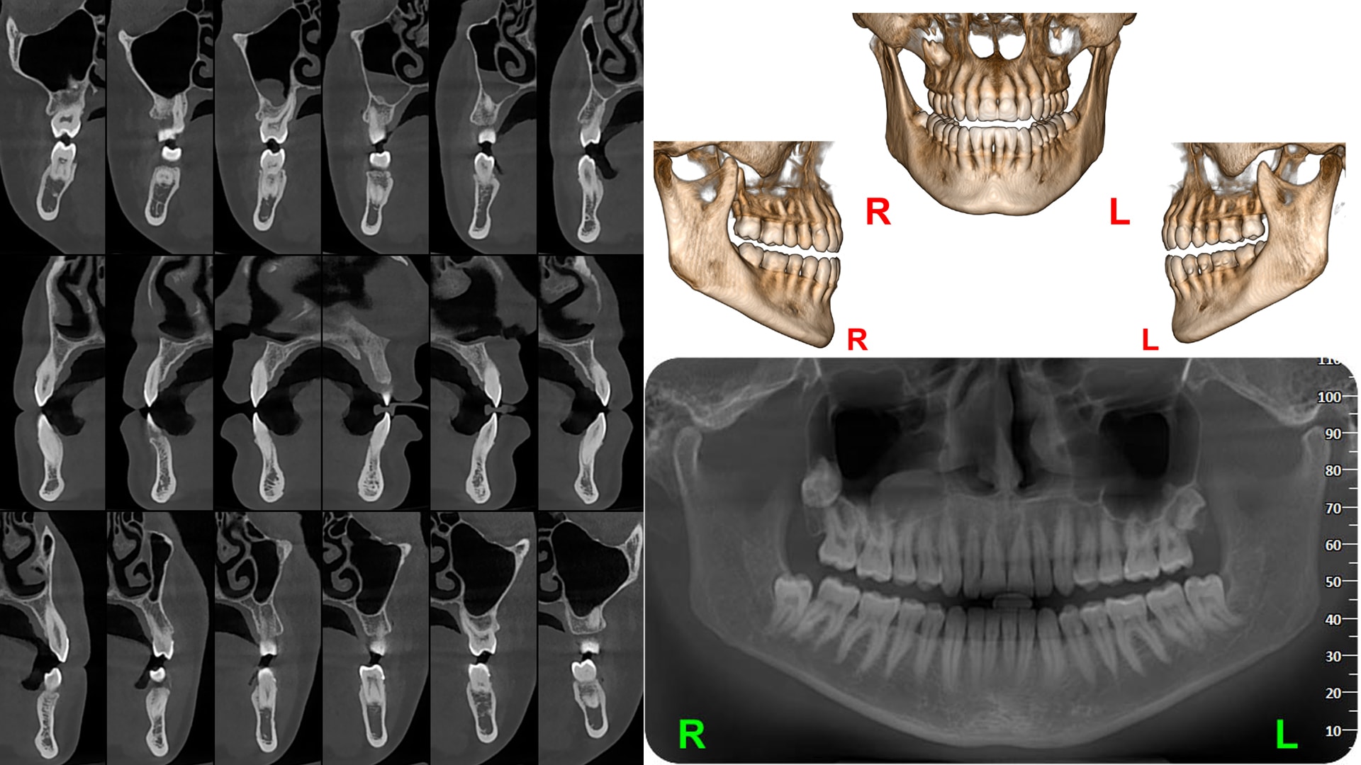
Complete tomography (maxilla + mandible) is a three-dimensional imaging technique used in dentistry, which provides accurate and detailed images of both arches, the zygomatic bone and the ascending mandibular ramus, providing information essential for diagnosis and treatment planning in dentistry.
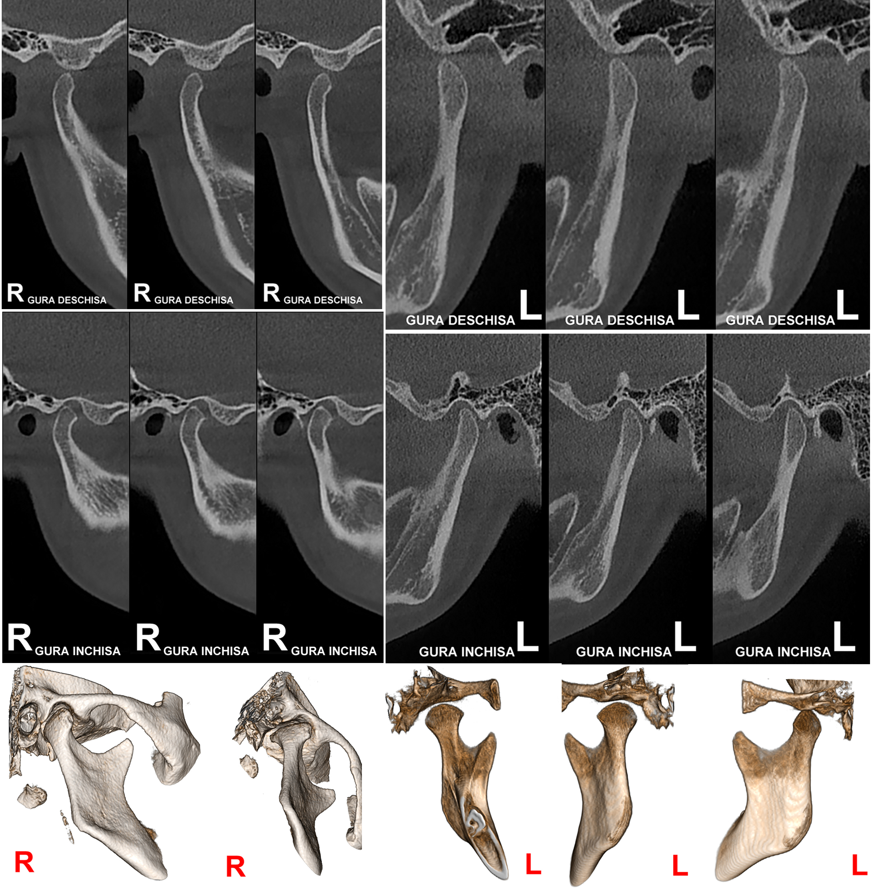
The CINE-X function represents the dynamic acquisition of stored radiological images through a video file. Movements can be visualized with this function present at the level of the Temporomandibular Joint during closing movements mouth opening respectively. The excursion can be visualized through the images captured from the lateral norm articular condyle at the level of the glenoid cavity. Images taken from the frontal norm allow the highlighting of possible asymmetries of the interincisal line trajectory over time opening-closing movements of the mouth.
The CINE-X function represents the dynamic acquisition of stored radiological images through a video file. Movements can be visualized with this function present at the level of the Temporomandibular Joint during closing movements mouth opening respectively. The excursion can be visualized through the images captured from the lateral norm articular condyle at the level of the glenoid cavity. Images taken from the frontal norm allow the highlighting of possible asymmetries of the interincisal line trajectory over time opening-closing movements of the mouth.
ATM tomography is a three-dimensional imaging technique used in dentistry, which provides precise and detailed images of the position of the condyles in the articular fossa both in the maximum intercuspation position and in the one with the mouth open, providing information essential for diagnosis and treatment planning in dentistry.
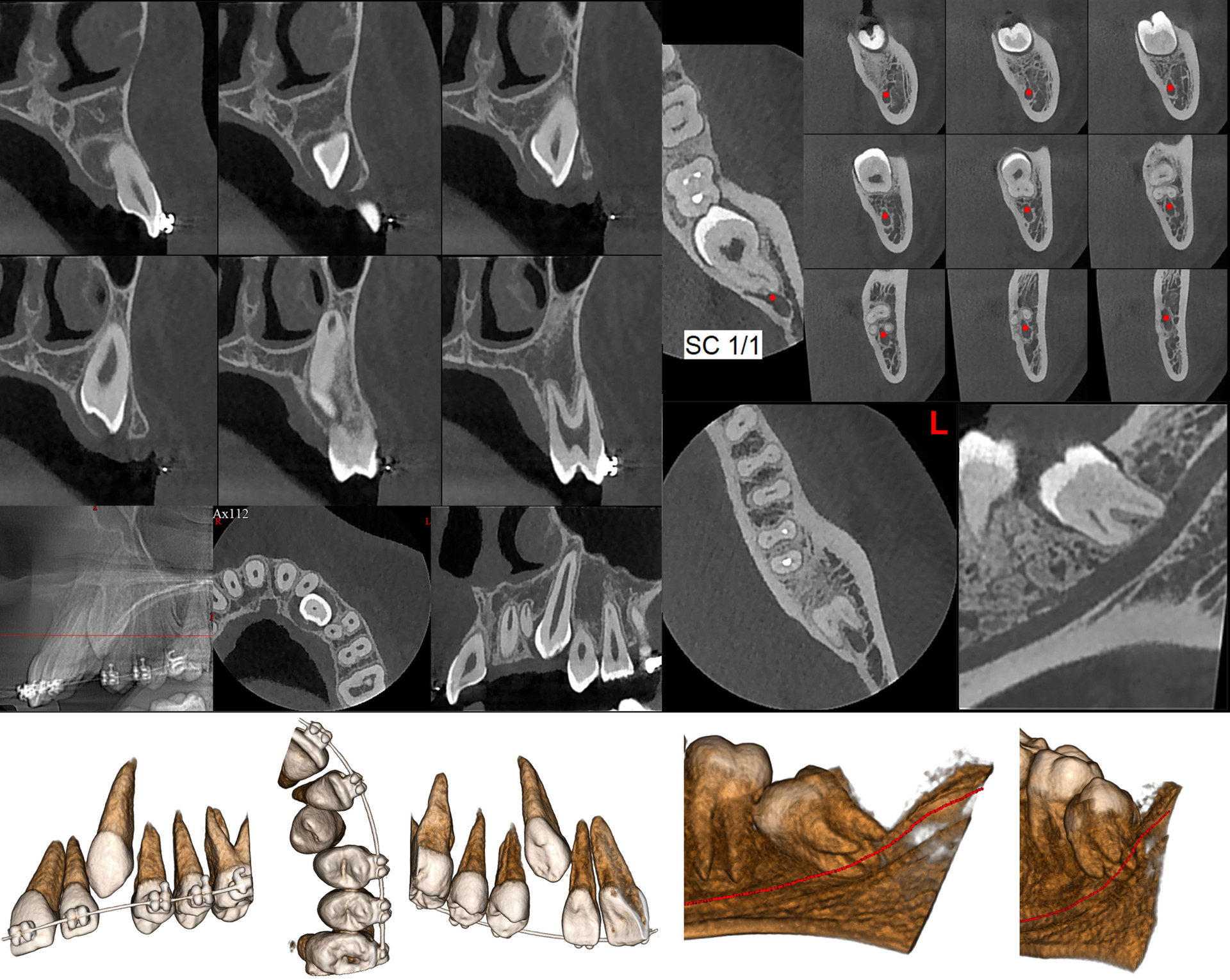
Tomography for dental abnormalities is a three-dimensional imaging technique used in dentistry, which provides accurate and detailed images of teeth and adjacent structures, being useful in the diagnosis and treatment planning for various dental anomalies, such as: anomalies of position, shape, number or alignment.
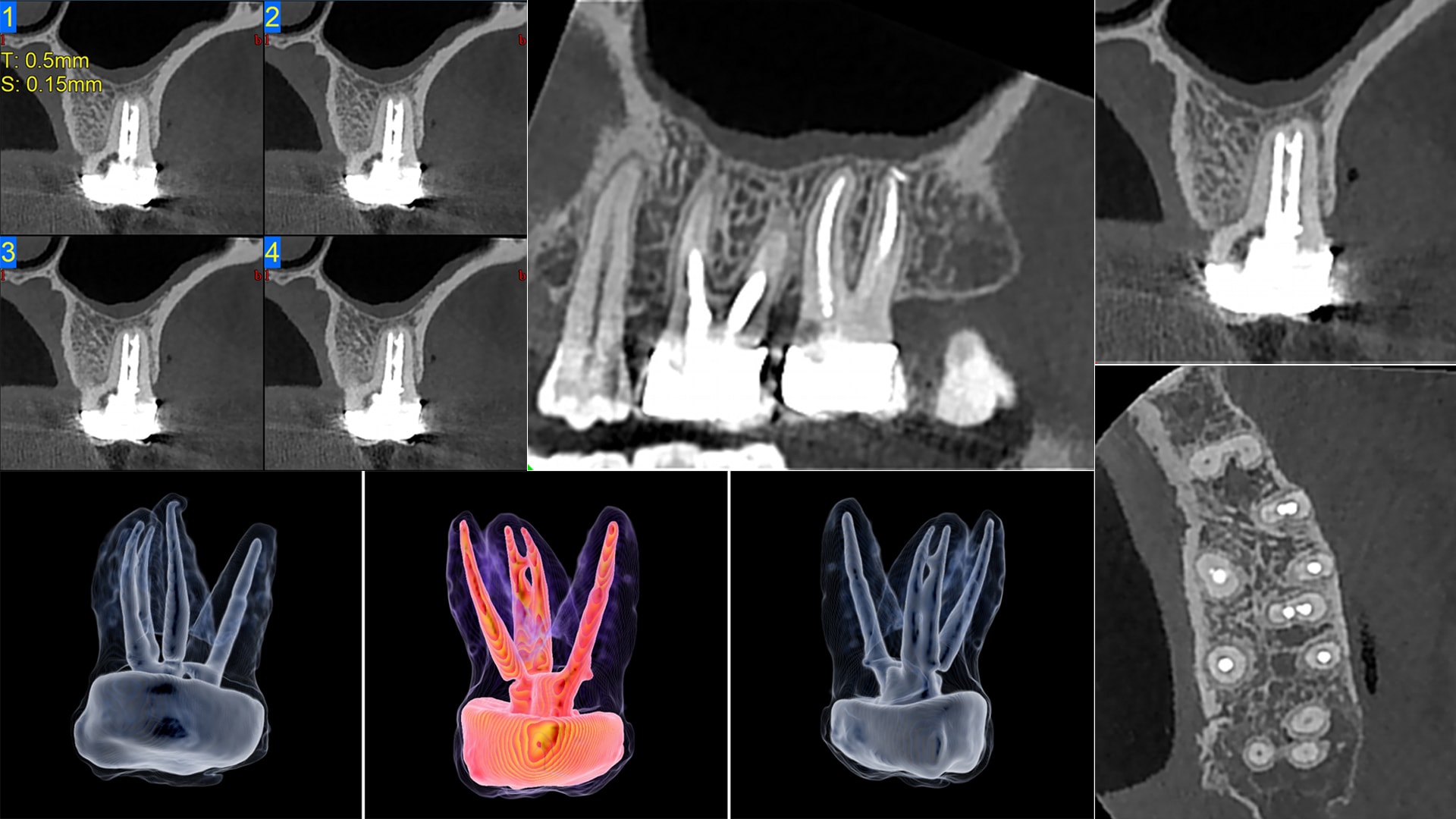
Tomography for endodontic purposes is a three-dimensional three-dimensional imaging technique detailed images of dental and periapical structures, helping to accurately diagnose and endodontic treatment planning, including identification of root canals, periapical lesions and associated complications.
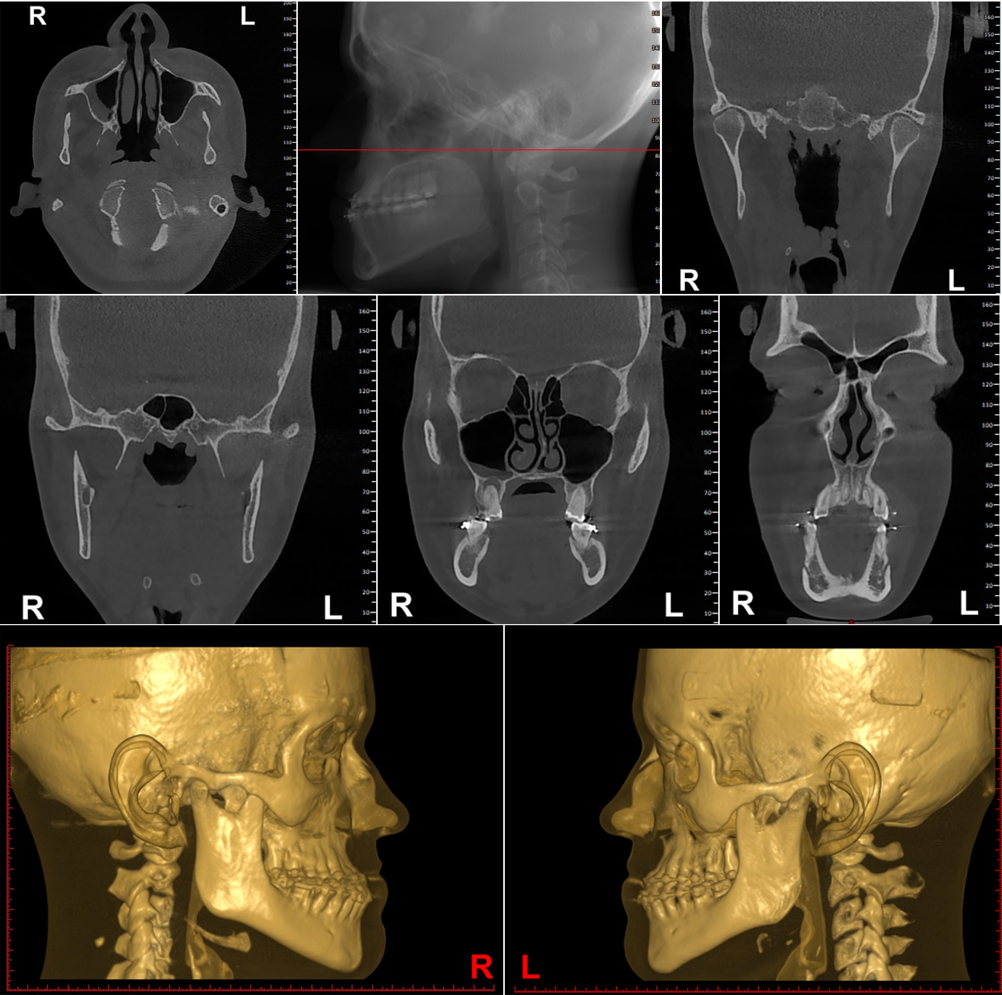
Tomography for orthognathic surgery provides detailed three-dimensional images of the craniofacial structures, helping the diagnosis and precise planning of interventions orthognathic surgery, including correct jaw repositioning and correction facial bone disharmonies.
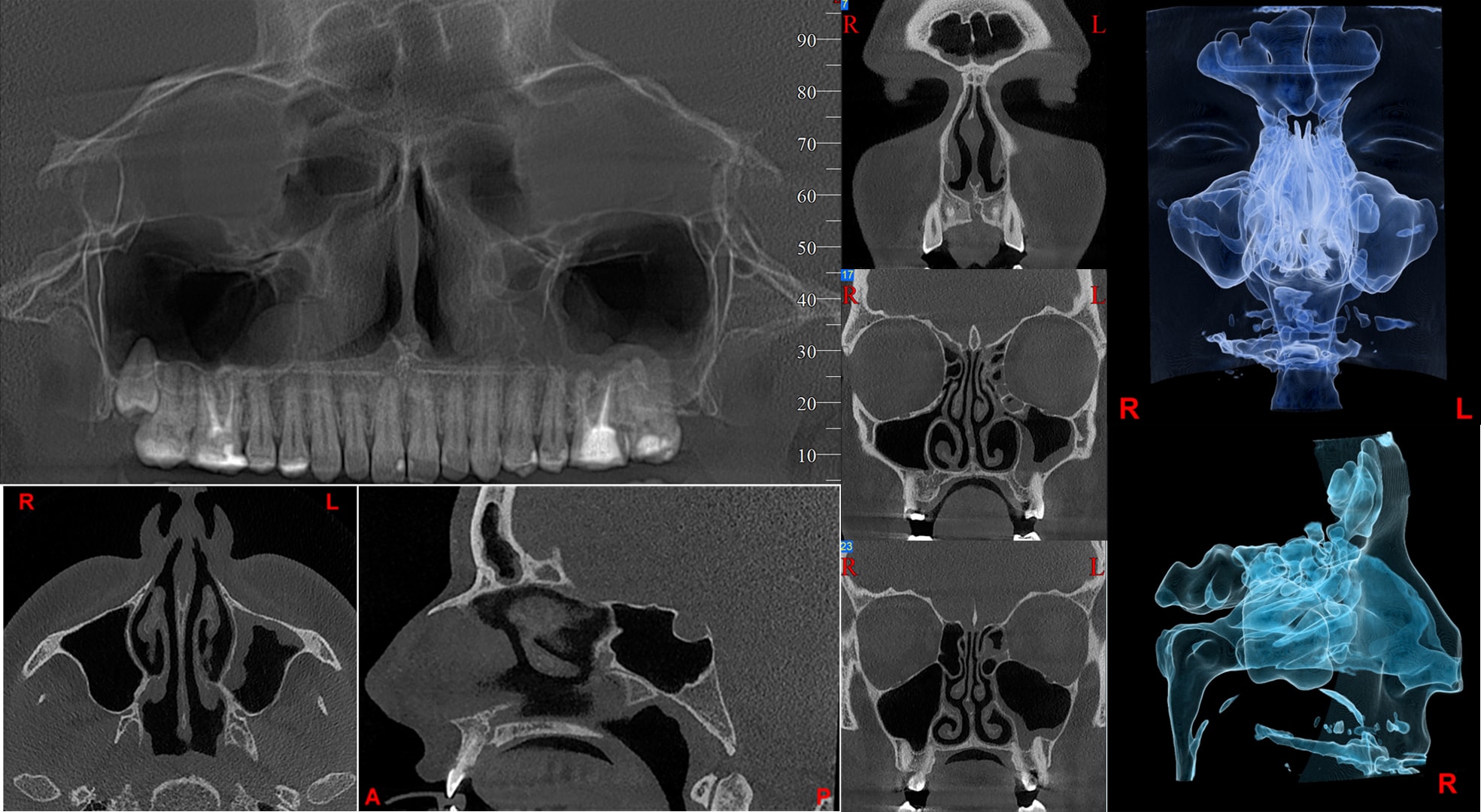
Tomography for the anterior sinuses of the face is a three-dimensional imaging technique which provides detailed images of the maxillary, paranasal, frontal and ethmoidal sinuses. It is used in the diagnosis and treatment planning for sinus conditions.


