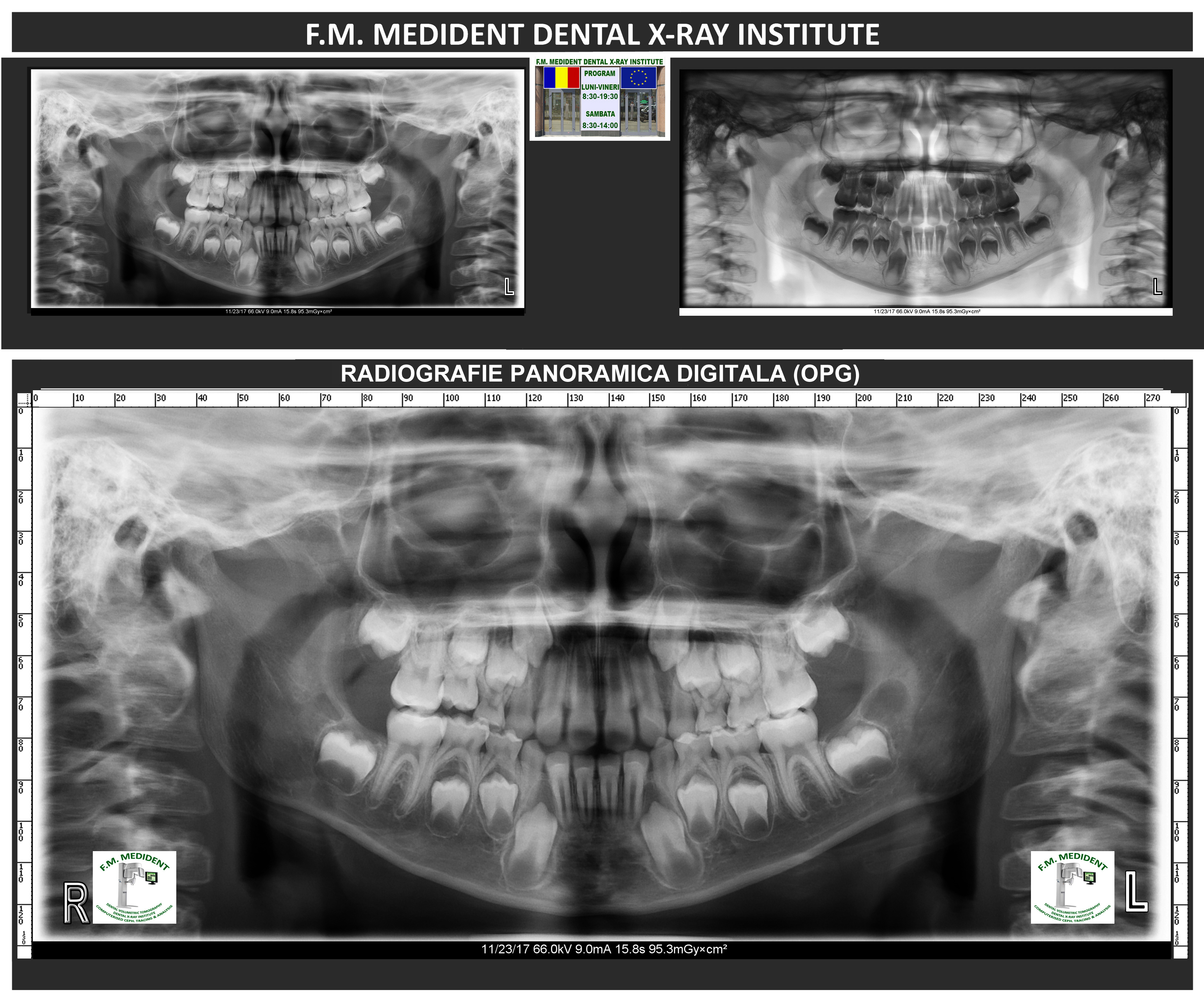
Panoramic radiograph shows the entire structure of the mouth providing information about present teeth, the alveolar process and the surrounding anatomical structures.
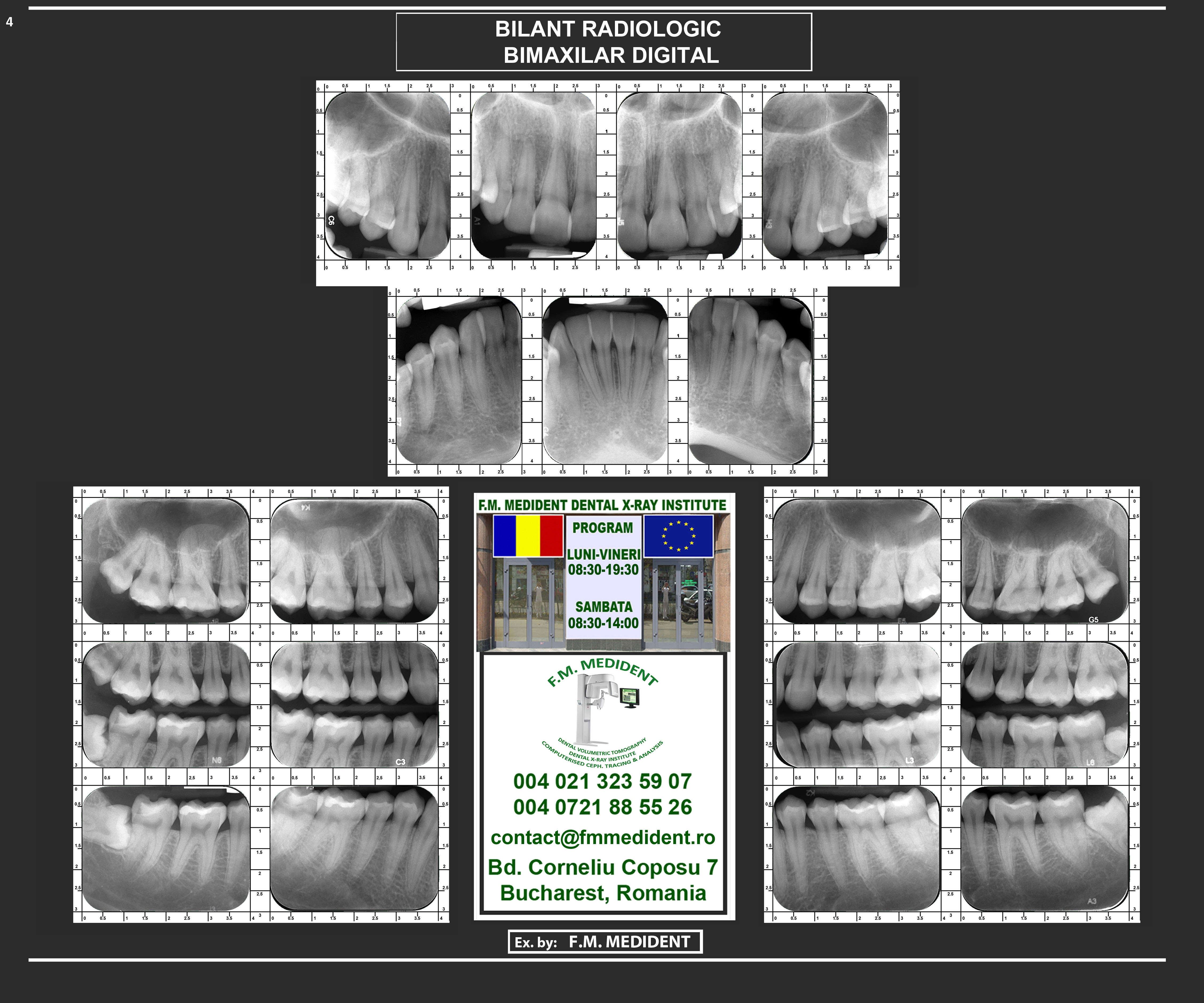
Includes both bitewing and periapical radiographs showing all present teeth on both upper and lower jaw.
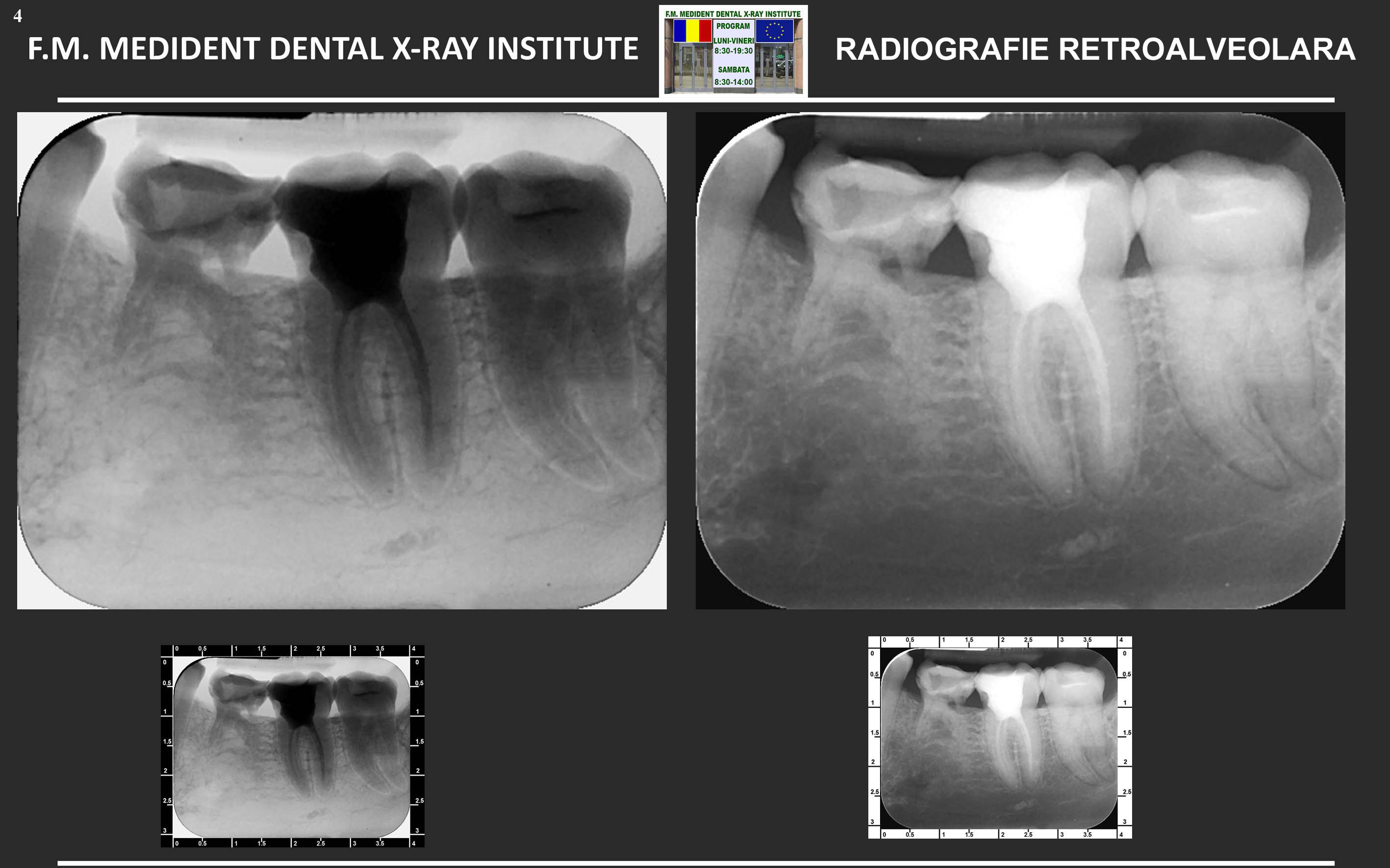
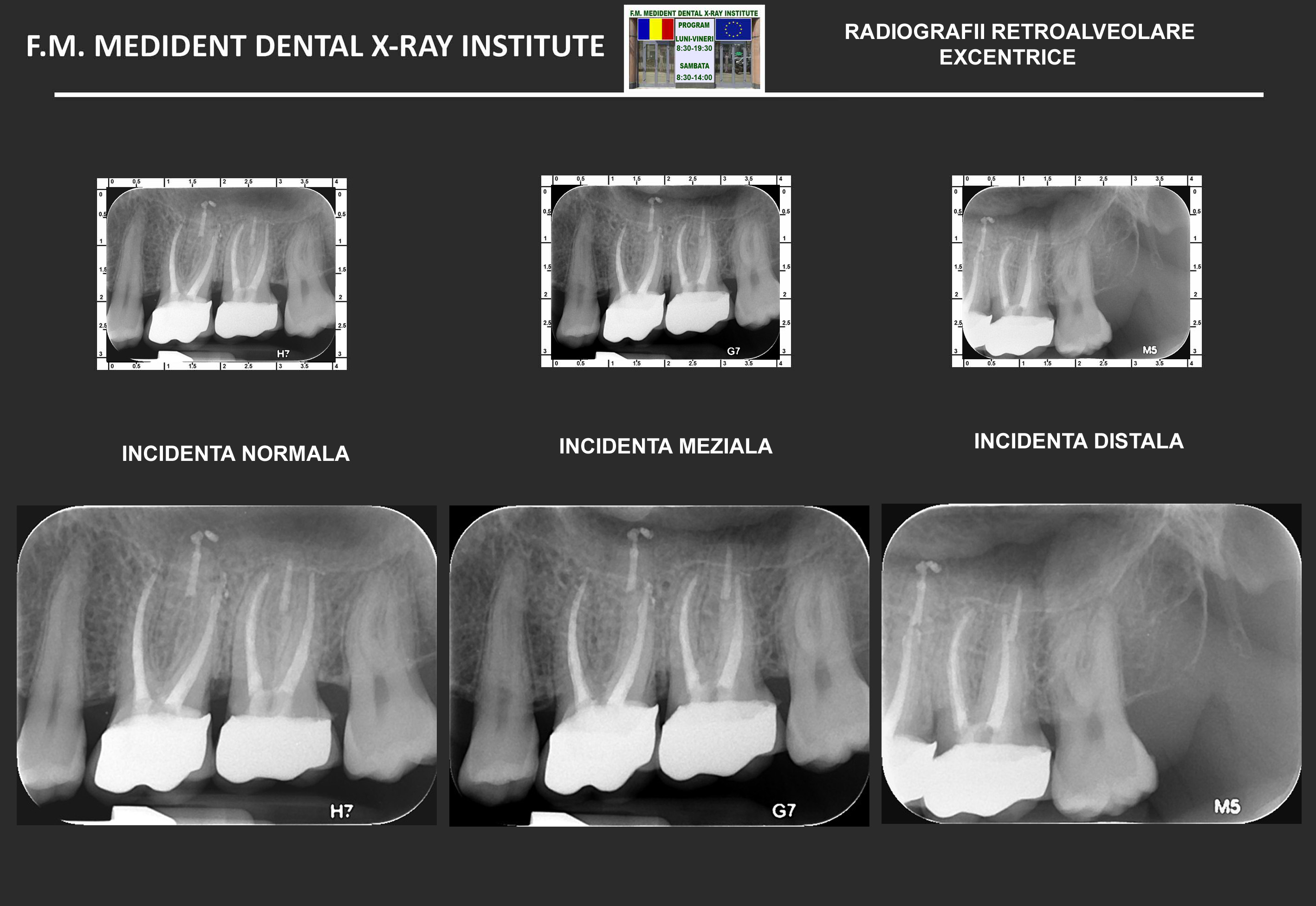
Periapical radiographs allow detailed visualization of both the crown and the root of the tooth. This type of radiograph is performed using the parallel technique (with a 90° angle to the long axis of the tooth) and focuses on a small area of 1–2 adjacent teeth.
Bitewing X-Rays show the crowns of the back teeth on each side of the mouth. With their help, interdental contact points can easily be observed.
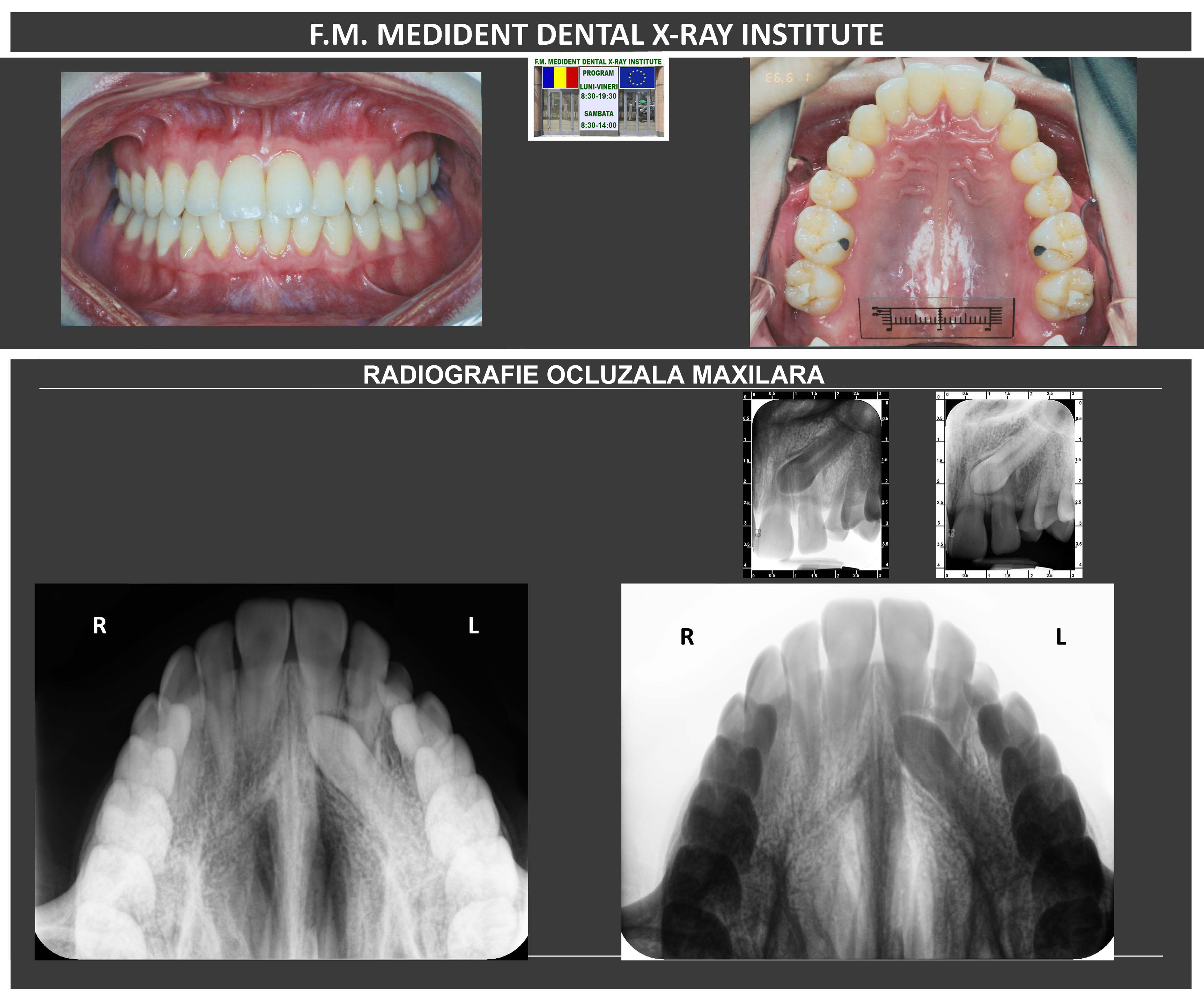
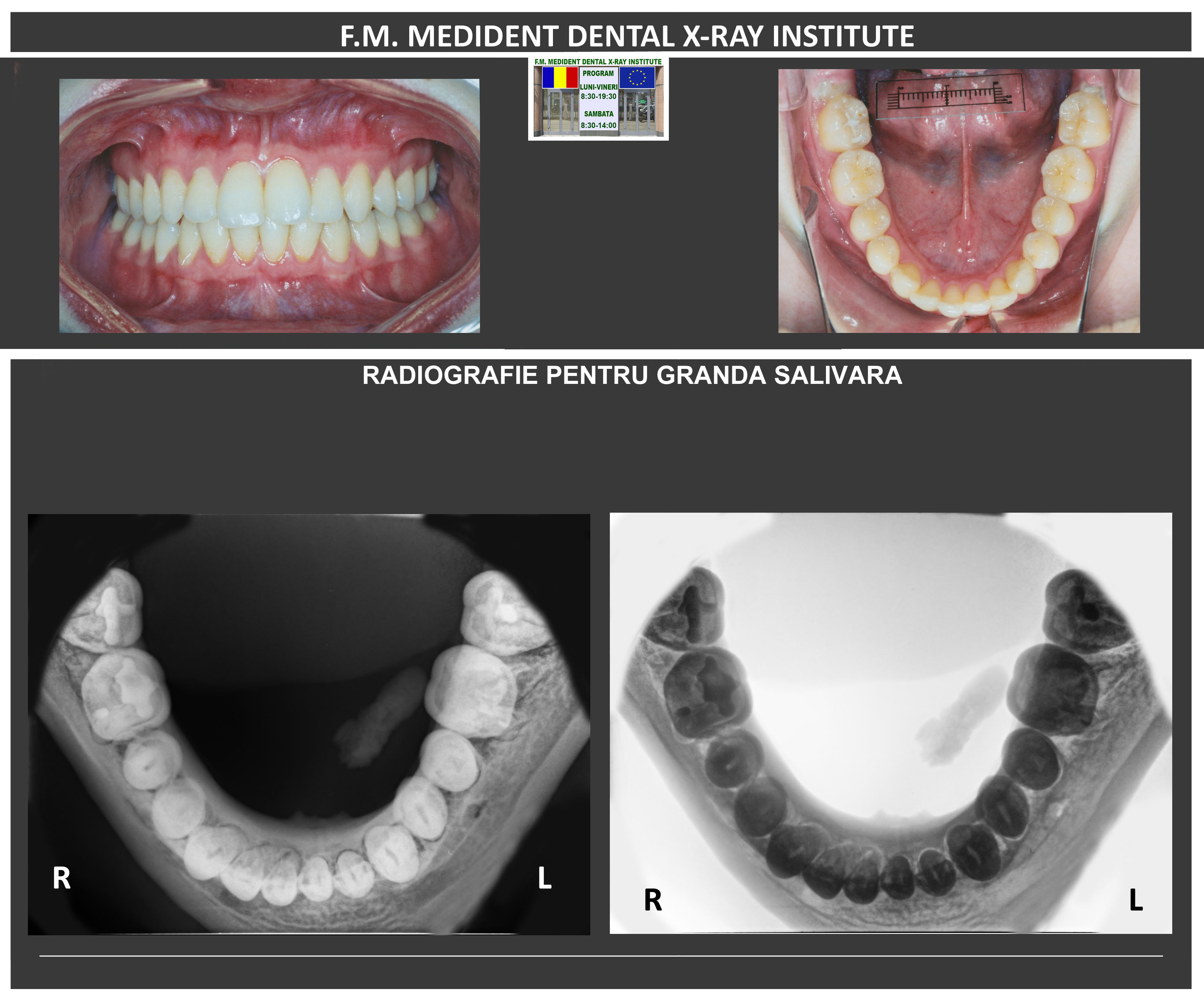
Occlusal radiography is the largest of the intraoral radiographs. This provides a cross-sectional view of the whole upper or lower jaw.
Salivary gland radiography aims to locate clavicles in the submandibular salivary glands and at the level of their channels. For the submandibular localization of lithiasis, simple x-ray radiography is performed with the bite film in the axial incidence of the buccal layer.
Eccentric periapical radiographies are composed of two periapical radiographs made from different angles (mesial and distal). These radiographs show their usefulness in endodontic treatment.
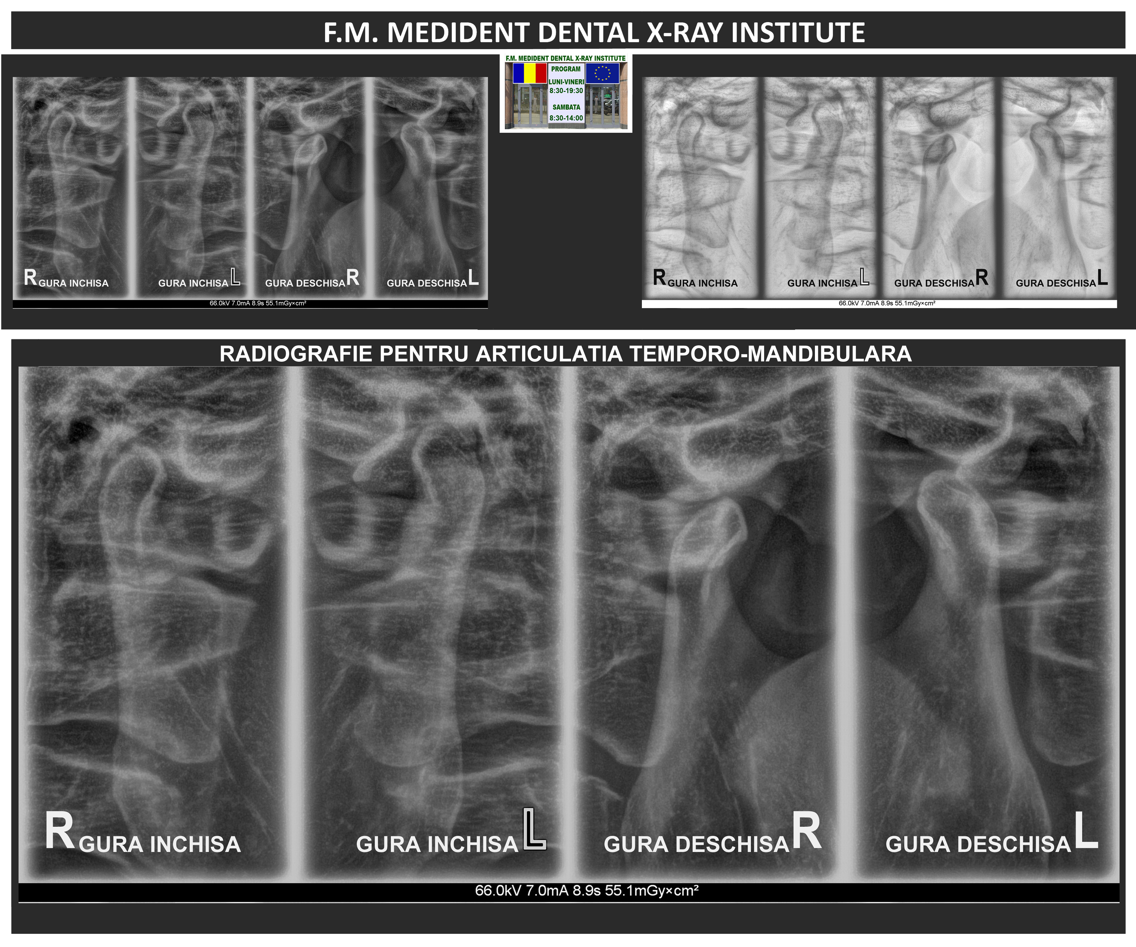
TMJ radiograph is composed of two static records of the condyle position in the glenoid fossa. The first recording is made in the maximum inter-positioning order (the closed mouth) and the second can be reached both in the maximum opening position and in the propulsion position.
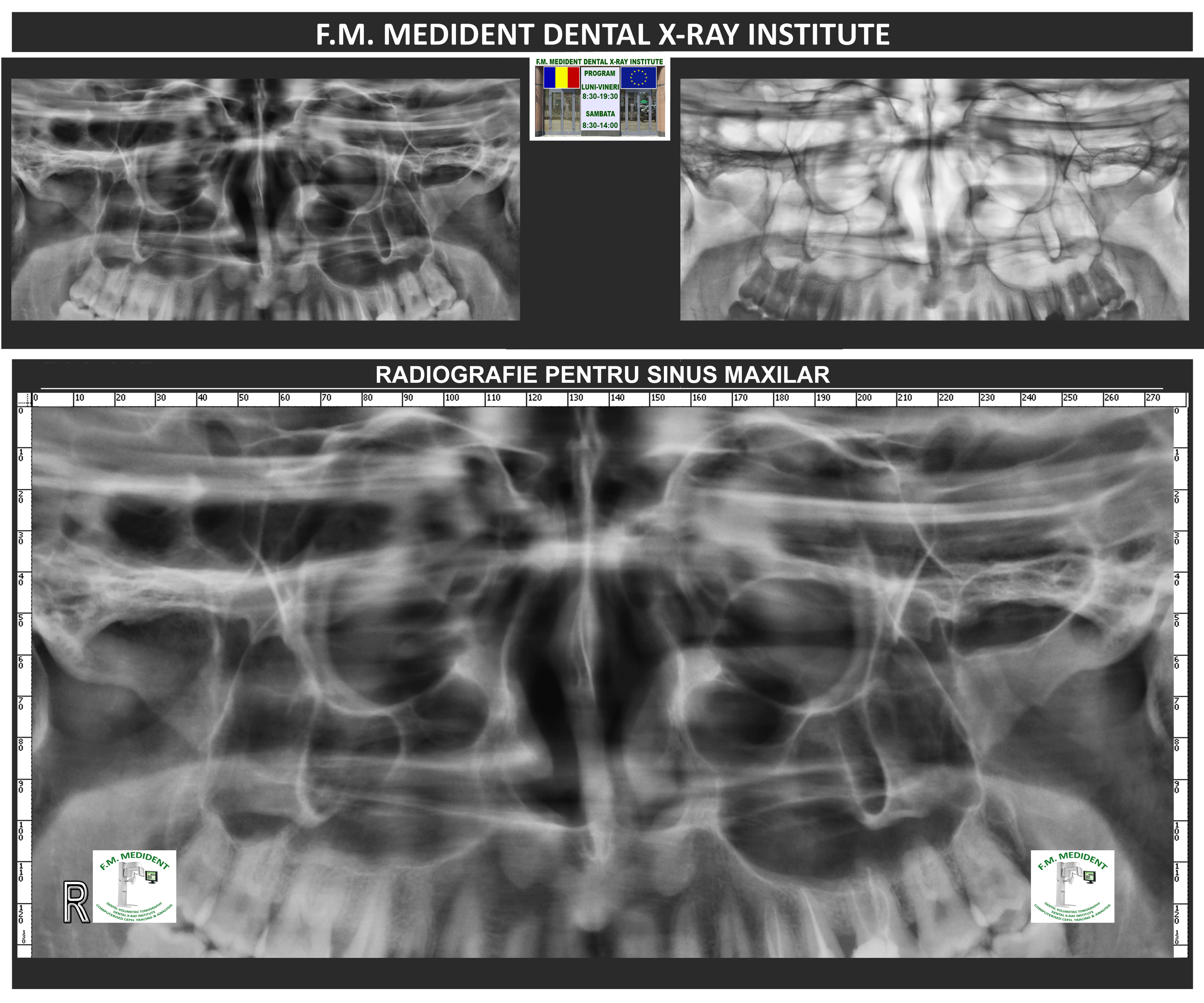
With this X-ray, you can clearly see the delimitations and shape of the sinus cavity and possible pathologies present in the maxillary sinuses.
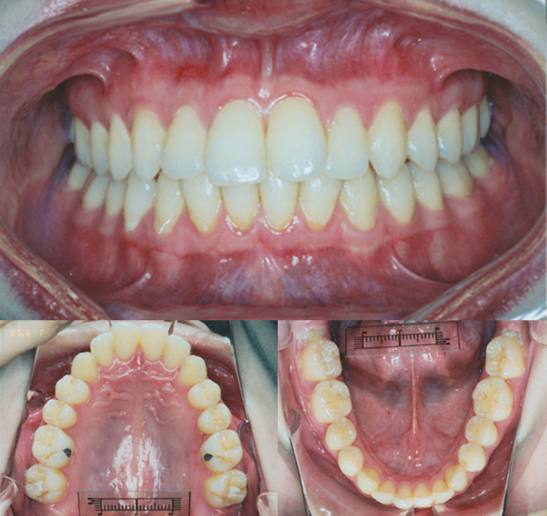
Diagnostic photographs are detailed images of teeth and gums taken in the oral cavity with a special camera, used for diagnosis, treatment evaluation and documentation of dental condition.


.jpg)